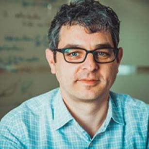
Thomas Heldt, associate director of IMES, and a core faculty member, describes how he closely collaborates with colleagues across MIT and at Boston-area hospitals, and how his research uses and analyzes physiologic data to aid clinical action.
Mindy Blodgett | IMES-HST
Thomas Heldt joined the MIT faculty in 2013 as a core member of the Institute for Medical Engineering and Science (IMES) and the Department of Electrical Engineering and Computer Science. Additionally, Thomas is a principal investigator with MIT’s Research Laboratory of Electronics (RLE). He directs the Integrative Neuromonitoring and Critical Care Informatics Group in IMES and RLE. He was recently named an associate director of IMES, where he will focus on internal affairs, among other duties. Heldt received his Medical Engineering and Medical Physics (MEMP) PhD from the Harvard-MIT Health Sciences and Technology (HST) program in 2004. Heldt's research interests include signal processing, estimation and identification of physiological systems, mathematical modeling, and model identification to support real-time clinical decision making, monitoring of disease progression, and titration of therapy, primarily in neurocritical and neonatal critical care.
Q.1 Your research focuses on signal processing, mechanistic mathematical modeling, and model identification. How does your research apply these technologies to solving clinical needs?
We look at current clinical environments and observe the volumes of multimodal physiologic waveform data that are collected on patients in critical care, peri-operative care or even emergency care. Much of this data is typically, visually, reviewed by the clinicians, and subsequently discarded after a holding period of just a few days, losing the opportunity for more systematic and patient specific analyses and insights. Critical to such analyses of these data streams is a deep understanding of the relevant physiology at the time scales of interest. We leverage insights from physiology, formulated in reduced order mathematical models, capturing the essential mechanisms that enable clinical action. We have applied this approach successfully to estimate intracranial pressure noninvasively, to make diagnostic decisions based on the analysis of the shape of the capnogram, and we are currently using ultrasound-based approaches to detect embolic events in patients on life support, such as ventricular assist devices or extracorporeal membrane oxygenation.
Q.2 You work closely with colleagues across MIT, and with clinicians at Boston-area hospitals, including Boston Children’s Hospital (where you hold a courtesy research appointment in neurology), Boston Medical Center (neurosurgery), and Massachusetts General Hospital (emergency medicine). What has been the fruit of some of these collaborations—what is the impact on your research?
Boston is a fantastic place to conduct translational research that crosses from our laboratories at MIT into the clinical environments for validation in the actual target patient population! The collaborative disposition and forward-thinking mindset of our clinician colleagues has really been fundamentally enabling for our research and has provided amazing mentoring to our students, postdocs, and me. We have collected validation data in brain-injured patients in the ICUs at Boston Medical Center and Children’s Hospital; we have collected pilot and validation data for our capnography work in the emergency departments at Children’s Hospital and Beth Israel Deaconess Medical Center; we have collected data for our emboli work in the operating rooms and ICUs at Children’s Hospital, and have analyzed the medical records of the neonatal ICU at Beth Israel Deaconess Medical Center and the emergency department at MGH.
Our work with the neonatologist at BIDMC, was focused on analyzing the monitoring alarm patterns in the neonatal ICU. We counted a staggering 177 alarms/baby/day, or one alarm every eight minutes on average, per baby. And this is a 54-bed neonatal ICU operating close to capacity every day! Such volumes of alarms contribute to noise pollution in an environment that should ideally be very calm. Additionally, since most of the alarms are nuisance alarms or do not require any clinical intervention, the clinical staff becomes desensitized to the alarm load and might end up ignoring truly important events. We analyzed the alarm patterns and alarm thresholds for a particular type of heart rate alarms and recommended a change in thresholds. This resulted in a 50% reduction of in-heart rate alarms per patient per day. Initially, the clinical staff had to file weekly reports to make sure the reduction in the alarm rate did not result in missed events. After about three months without a single reportable event, the hospital safety committee approved the change.
With colleagues from the MGH Department of Emergency Medicine, we developed and tested a triage rule to identify patients at risk of septic shock. At the time, the MGH ED saw about >120,000 patients/year, and around 75% of patients, who ended up in the ICU with severe sepsis and septic shock came through the emergency department. Hence, ED triage was the first point of patient contact and the first opportunity to flag patients for possible sepsis and septic shock, and initiation of goal directed therapy. One result of our work was a significant reduction in the time to appropriate antibiotic administration. The work was subsequently validated in other Partners hospitals and implemented in the electronic medical record system of Partners-affiliated hospitals.
Q.3 Can you tell us a bit about your background, and about how it is that you became interested in systems-physiology and biomedicine? What are your goals for your research, and for your career?
That is a longer story! In short, I started out studying physics back in Germany. After a while, I got interested in applying concepts I learned in physics to physiology and medicine, so I designed my own MD/PhD program by picking up medicine as a second major. Through some fortuitous events, I ended up attending surgeries for congenital heart defects for about a term. This was a very formative experience and almost pushed me toward dropping physics and going all out on becoming a surgeon. However, I had also always wanted to spend part of my education abroad and had applied to various universities in the US. I ended up getting admitted to the graduate physics program at Yale and spent a couple of years doing nonlinear optics. While I loved the work at Yale and had a fantastic mentor, I missed the clinical exposure and application of my work to medicine. I had heard about the HST program and decided to send in an application. I joined the MEMP program in 1997 and have been at MIT ever since.
In our current research, we are very interested in providing better monitoring modalities for patients with brain injuries. We are developing novel algorithmic and device approaches so we can replace the current invasive monitoring modalities with entirely noninvasive ones, and provide additional clinically actionable information that gives insights on the physiology of the injured brain, and can help guide treatment decisions. I would like to see some of these technologies through to routine deployment at the bedside.
The great thing about being in IMES and MIT is that everybody is very collaborative. What I am looking forward to is much of the same, working with colleagues in IMES, on important problems that maybe none of us are able to tackle alone, but that together we have a real chance of solving—and having fun along the way!
